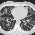
CMV Pneumonia on CXR
Case courtesy of our radiology website partner, RiT.Axial CT image of the same patient confirmed a nodule...
Hangman’s Fracture (X-ray)
Image courtesy of our radiology website partner, RiT.Lateral view of a cervical spine...
Hangman’s Fracture (CT)
Image courtesy of our radiology website partner, RiT.Axial CT image of the cervical spine at C2 level...
Pelvic Congestion Syndrome (CT)
Case courtesy of our radiology website partner, RiT.Axial CT image of a 42-year-old woman with chronic pelvic...
Candida Esophagitis
Case courtesy of our radiology website partner, RiT.Double contrast barium esophagography shows innumerable pseudomembranes and plaques (arrows)...
Crazy Paving on Chest CT
Crazy Paving is a appearance that is basically a linear network or reticular pattern in the crazy-paving...
Cardiac MRI Nonimpaction
Noncompaction - Four-chamber - DE-MRI reveals hypertrabeculated noncompacted myocardium in the apex and distal portions of the...
Cardiac MRI – Arrhythmogenic Right Ventricular Dysplasia (ARVD)
ARVD. Right-ventricular horizontal long-axis DE-MRI demonstrates diffuse, subendocardial enhancement of thinned RV wall, consistent with fibrous replacement...
Cardiac Sarcoidosis – Cardiac MRI
Cardiac sarcoidosis. Mid ventricular short-axis DE-MRI demonstrates patchy, mid myocardial scarring in the...
Pericarditis – Cardiac MRI
Pericarditis. 2-chamber DE-MRI demonstrates diffuse, intense pericardial enhancement, more prominent inferiorly, consistent with...


