Chronic PE – Chronic Pulmonary Embolism
Case submitted/via our partner RiT.Axial CT image shows a partial thrombus (stars) in the main and proximal...
SEGA MRI – Subependymal Giant Cell Astrocytoma
SEGA MRI - Subependymal Giant Cell Astrocytoma. Axial and coronal MR images (contrast enhanced T1WI) show an...
Lipohemarthrosis – Sagittal CT
Lipohemarthrosis* Mixture of blood and fat in a joint cavity following trauma* Fat from the marrow space...
Lipohemarthrosis – Cross-table lateral radiograph
Lipohemarthrosis* Mixture of blood and fat in a joint cavity following trauma* Fat from the marrow space...
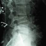
Lumbar Compression Fracture (X-Ray)
Diagnosis: Compression fracture of the fourth lumbar vertebra post falling from a height.
A...
Leptomeningeal Carcinomatosis
Sagital T1 (post contrast) MR image shows nodular/irregular enhancement of the leptomeninges, especially in the posterior fossa (arrows). The patient was...
Pericardial Effusion
Description of above image:
Lateral chest radiograph shows small bilateral pleural effusions....
Bucket Handle Meniscal Tear on Knee MRI
Fig: Coronal T2 image (with fat suppression) shows a displaced fragment (arrow) of medial meniscus into the...
Pellegrini-Stieda Lesion on Knee X-Ray
Diagnosis: Pellegrini-Stieda Lesion on Knee X-Ray
Discussion:
Pellegrini-Stieda lesions are named...
Posterior Shoulder Dislocation with Reverse Hill-Sachs lesion
Key Points:
Posterior shoulder dislocations (PSD) are most likely associated with reverse Hill-Sachs lesion...


