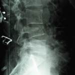
Lipohemarthrosis – Cross-table lateral radiograph
Lipohemarthrosis* Mixture of blood and fat in a joint cavity following trauma* Fat from the marrow space...
Lumbar Compression Fracture (X-Ray)
Diagnosis: Compression fracture of the fourth lumbar vertebra post falling from a height.
A...
Leptomeningeal Carcinomatosis
Sagital T1 (post contrast) MR image shows nodular/irregular enhancement of the leptomeninges, especially in the posterior fossa (arrows). The patient was...
Pericardial Effusion
Description of above image:
Lateral chest radiograph shows small bilateral pleural effusions....
Pellegrini-Stieda Lesion on Knee X-Ray
Diagnosis: Pellegrini-Stieda Lesion on Knee X-Ray
Discussion:
Pellegrini-Stieda lesions are named...
Posterior Shoulder Dislocation with Reverse Hill-Sachs lesion
Key Points:
Posterior shoulder dislocations (PSD) are most likely associated with reverse Hill-Sachs lesion...
Lung Abscess (Radiograph)
Key Points:
A primary lung abscess develops usually as a result of an infection...
Lumbar Vertebral Fracture
This fracture shown here is a compression type vertebral body fracture that may often appear as wedge...
Esophageal Varices
Diagnosis: Esophageal Varices (Fluoroscopy)
Discussion:
Esophageal varices are dilated...
Lymphocytic Interstitial Pneumonia (LIP) from Amyloidosis
Case Quick Facts:


