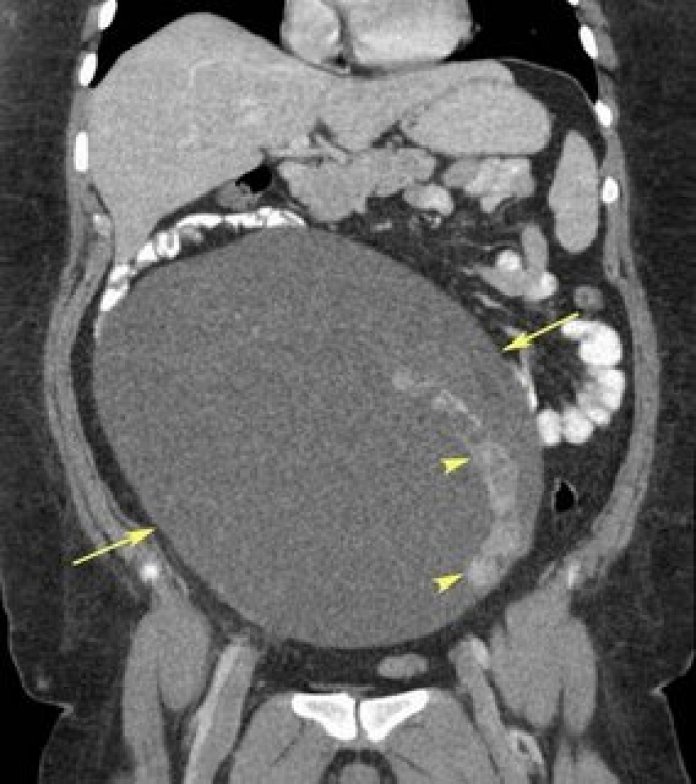
Functioning Ovarian Tumor contributed via our partner RiT.
Coronal reformatted CT image shows a very large mixed cystic/solid mass in the abdomen/pelvis of a 25-year-old woman presenting with hirsutism. On CT, her normal right ovary is not visualized. The left ovary is normal.
Facts
* Several ovarian tumors and tumorlike conditions can produce estrogen or androgen, resulting in signs and symptoms of hyperestrogenism or hyperandrogenism
* Enlarged uterus with thick endometrium can be seen on imaging in patients with hyperestrogenism
Tumors/tumorlike conditions producing hyperandrogenism
* Sertoli-Leydig cell tumor
* Leydig cell tumor
* Gynandoblastoma
* Germ cell tumor (carcinoid)
* Brenner tumor
* Tumorlike conditions (polycystic ovary syndrome, stromal hyperplasia, stromal hyperthecosis, hyperreactio luteinalis, pregnant luteoma)
Tumors producing hyperestrogenism
* Granulosa cell tumor
* Thecoma
* Serous epithelial tumor
* Mucinous epithelial tumor
* Endometrioid tumor
Tumors that can produce either estrogen or androgen
* Metastatic tumor
* Stromal luteoma
* Sclerosing stromal tumor
Our case: Carcinoid tumor of the right ovary.
Reference:
Tanaka YO, Tsunoda H, Kitagawa Y, et al. Functioning ovarian tumors: direct and indirect findings at MR imaging. Radiographics 2004;24:S147-S166.


