
Diagnosis: Gastric carcinoma
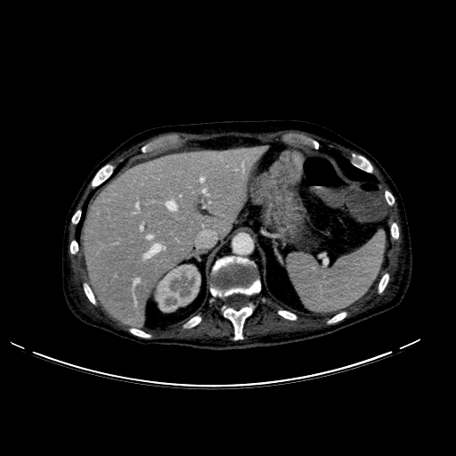
Findings: Mass is seen along the medial border of the stomach wall extending towards the liver. Disrupted ruggae are partially visualized. No ulceration is identified clearly on this image but given anemia, it is possible CT may not visualize.
Key points: On CT, look for polypoid mass with or without ulceration. Focal wall thickening can also be seen. Infiltration and mucosal irregularity may be possible. Loss of normal rugal fold pattern is also another sign seen on imaging. Accurate staging would enable surgical and/or medical treatment decisions to maximize positive clinical outcomes.
Credit: Case submitted by Dr. Giuseppe Pelle MD to radRounds Radiology Network


")

